Figure 2.
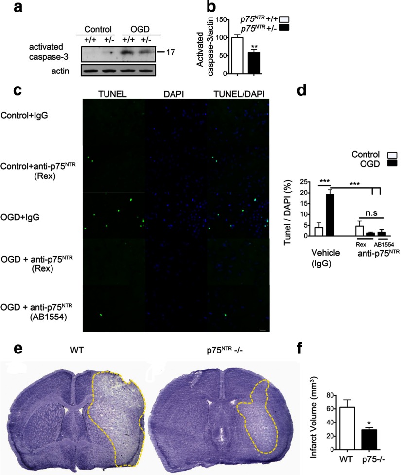
Blockade of p75NTR rescues ischemic cell death. a, Organotypic hippocampal slices from p75NTR haploinsufficient mice and wild-type littermates were subjected to 30 min of OGD and harvested at 8–12 h after OGD. OGD-induced activated caspase-3 was significantly decreased in hippocampal slices from p75NTR haploinsufficient mice. b, Quantification of activated caspase-3 after OGD. Values are shown relative to activated caspase-3 in the wild type (n = 3; Student's t test). c, Apoptotic cell death is significantly reduced in the presence of p75NTR function-blocking antibodies. Primary hippocampal neurons were treated with anti-p75NTR antibody (REX) or AB1554 (Millipore). Control cells were treated with rabbit IgG (Millipore). Antibodies were added 2 h before OGD, and TUNEL assay was performed 24 h after OGD. Representative images show a decrease in OGD-induced apoptosis on blockade of p75NTR. Scale bar, 20 μm. d, Quantification of TUNEL-positive cells represented as a percentage of DAPI cells in the field. A total of three fields were imaged from each coverslip (n = 4). ANOVA analysis was performed. e, Representative cresyl violet-stained coronal brain sections from wild-type and p75NTR−/− mice 72 h after MCAO. Note the smaller infarct (unstained white area) in p75NTR−/− mice. f, Quantification of the infarct volume 72 h after MCAO in wild-type (n = 7) and p75NTR−/− (n = 5) mice shows a significant infarct reduction in p75NTR−/− mice. *p < 0.05; **p < 0.01; ***p < 0.001, Student's t test.

