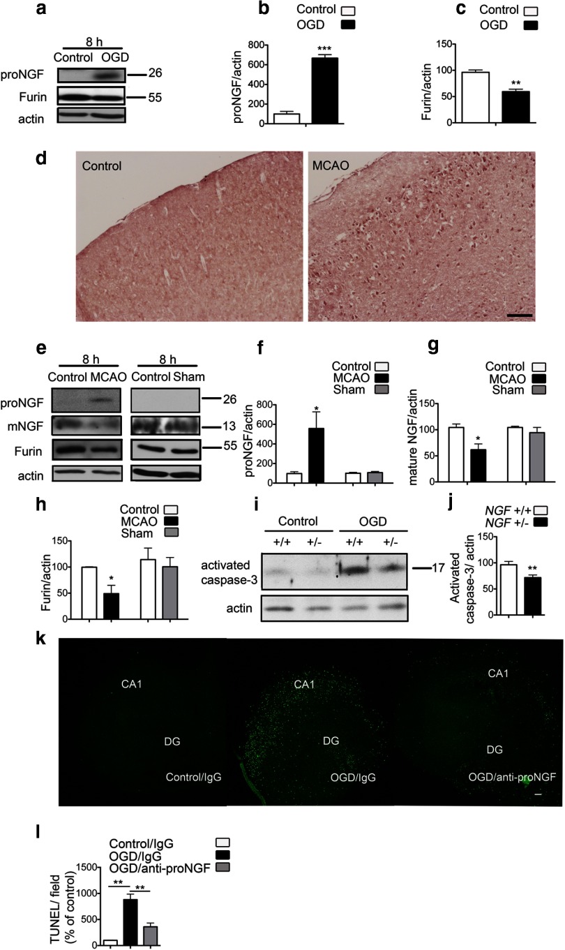
Figure 3.
ProNGF is increased after ischemia. a, ProNGF protein expression is increased with a concurrent decrease in furin in organotypic hippocampal slices subjected to OGD. b, Quantification of proNGF levels from a (n = 3; Student's t test). c, Quantification of furin levels from a (n = 3; Student's t test). d, Immunohistochemistry using an anti-pro-domain antibody shows induction of proNGF protein in the ischemic cortex 8 h after MCAO, compared with the contralateral control hemisphere. Images are representative of data from three different animals. Scale bar, 100 μm. e, Immunoblots from the ischemic cortex show elevation of proNGF after MCAO. Mature NGF is decreased in the ischemic cortex. A concomitant decrease in furin is also observed in the ischemic hemisphere. Sham surgeries do not affect the levels of proNGF, NGF, or furin expression. f, Quantification of proNGF levels from e (n = 4). Student's t test was used to compare between the MCAO or sham groups with the contralateral control hemisphere. g, Quantification of mature NGF from e (n = 3; Student's t test). h, Quantification of furin levels from e (n = 4). i, OGD-induced activation of caspase-3 is decreased in slices from ngf +/− mice compared with slices from wild-type littermates. j, Quantification of activated caspase-3 from i. Values are shown relative to activated caspase-3 in the wild type 8–12 h after OGD (n = 3; Student's t test). k, Anti-proNGF treatment in organotypic hippocampal slices rescues OGD-induced apoptotic cell death. The function-blocking antibody specific for the pro-domain of proNGF or nonimmune rabbit IgG was added to slices 2 h before OGD, and TUNEL assay was performed at 24 h after OGD. Representative images of slices are shown. DG, Dentate gyrus. Scale bar, 100 μm. I, Quantification of TUNEL positivity per field shows significant reduction in apoptosis on treatment with the anti-proNGF antibody. Three to five slices in each group were analyzed from four independent experiments (n = 4; ANOVA). *p < 0.05; **p < 0.01; ***p < 0.001.

