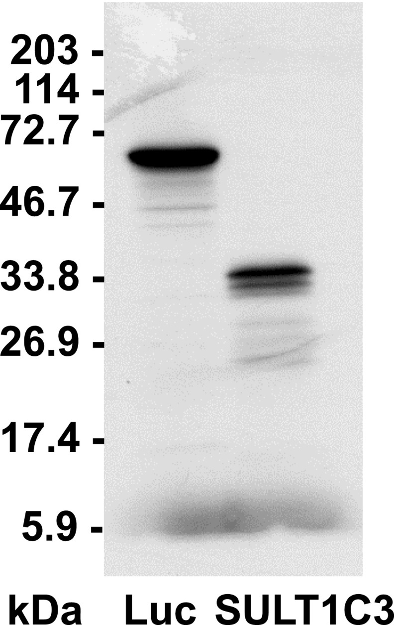
Fig. 5.
In vitro transcription/translation of a SULT1C3 cDNA clone containing exons 7a and 8a. A plasmid (pcDNA3.1) containing SULT1C3 cDNA that had been amplified from LS180 cells, from 4 nt downstream of the transcription start site indicated by 5′-RACE to 127 nt downstream of the translation stop codon in exon 8a, was analyzed by in vitro transcription/translation in a rabbit reticulocyte system. After in vitro transcription/translation, proteins were separated by SDS-PAGE and 35S-labeled SULT1C3 was visualized by fluorography. Lane one shows in vitro transcription/translation of a positive control luciferase DNA (Luc, ∼61 kDa). Locations of molecular weight size markers are indicated to the left of the image.

