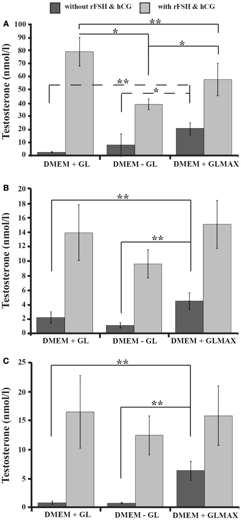
Figure 2.
The influence of different DMEM culture media on testosterone production by the Leydig cells in three-dimensional cultures of rat testicular cell. The cells were cultured for 14 days in DMEM + glutamine (GL), DMEM − glutamine (GL), or DMEM + Glutamax (GLMAX) (presented on the X-axis) either with (light columns) or without (dark columns) hCG and rFSH stimulation. The concentration of testosterone in the culture medium following 1 day (A), 7 days (B), and 14 days (C) of culture (determined by radioimmunoassay and expressed in nanomoles/liter) is shown on the Y-axis. One-way RM ANOVA with the Shapio–Wilk test for normality was applied to compare the different experimental conditions. *p < 0.05,**p < 0.01.

