Figure 4.
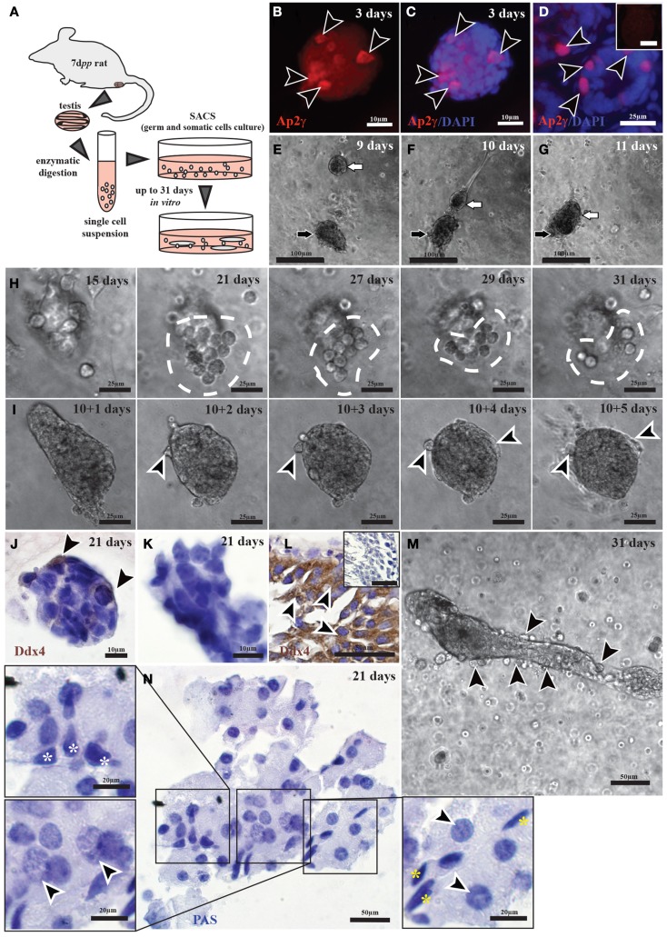
Tubule formation by and germ cell differentiation of pre-pubertal rat testicular cells in three-dimensional cultures. (A) A schematic overview of the experimental conditions. (B–D) Immunofluorescent staining of undifferentiated spermatogonia (Ap-2γ) in cell colonies originated from culturing cells from 7-day-old rats for 3 days (B,C), as well as from 8-day-old rats as a positive control (D) [Ap2γ: red staining (black arrow heads); DAPI: blue staining]. The negative control with IgGs is shown as small insert in (D). (E–G) 9- (E), 10- (F), and 11-day cultures (G) showing two colonies (the black and the white arrows) migrating toward one another. (H,I,M) Active migration of cells out of colonies cultured for as long as 31 days [white dashed line in (H), black arrows heads in (M)] or following incubation of isolated cell colonies in liquid medium for 5 days [black arrow heads in (I)]. (J–L) Immunohistochemical staining for germ cells (Ddx4) in colonies originating from 7-day-old rats and cultured for 21 days (J,K), as well as from 60-day-old rat testis [positive control; (L)] [Ddx4: brown staining (black arrow heads); Hematoxylin: blue staining]. (N) Cells cultured for 21 days exhibit morphologies similar to those of peritubular cells (yellow stars), Sertoli cells (white stars), leptotene spermatocytes, and early pachytene spermatocytes (black arrow heads).

