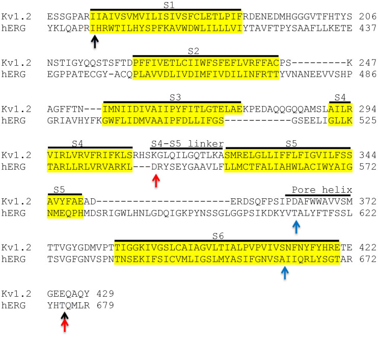
Figure 1. Sequence alignment of Kv1.2 and hERG showing sites of substitutions.
Potential transmembrane regions (highlighted in yellow) and other functional elements (S4–S5 linker and pore-helix) are labelled. Arrows indicate sites where different chimaeras were joined: Black, S1–S6; red, S4/S5–S6; blue, P-S6.

