Figure 1.

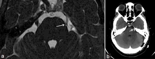
MRI brain (CISS sequence) depicting small vessel (arrow) at the trigeminal nerve juxtapontine segment (a). Post-MVD CT brain demonstrating the Teflon patch (arrow) at the juxtapontine and cisternal segments of the trigeminal nerve (b)

MRI brain (CISS sequence) depicting small vessel (arrow) at the trigeminal nerve juxtapontine segment (a). Post-MVD CT brain demonstrating the Teflon patch (arrow) at the juxtapontine and cisternal segments of the trigeminal nerve (b)