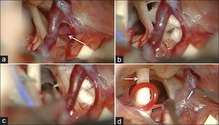
Figure 3.

Intraoperative photos demonstrating the left trigeminal nerve and artery loop at the upper surface of the juxtapontine nerve segment (arrow) (a). The artery loop was isolated from the nerve using Teflon patch (b). Note the trigeminal nerve atrophy, flattening and changes in color as compared with the faciocochlear complex (arrow) (a, b and d). The site of trigeminal nerve biopsy at the midcisternal segment is demonstrated (c)
