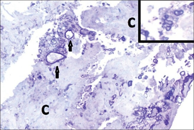
Figure 4.

Histopathology thick section of the specimen showing abundant collagen fibers (C) where nerve sprouts are embedded. In the center two large and thinly myelinated axons (arrows) have empty axoplasm. Inset: A regenerating axonal cluster. (Toluidine blue. Original magnifications × 400)
