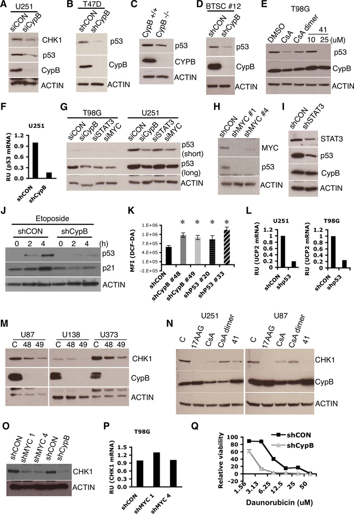
Figure 5. Cyclophilin B regulates p53 and Chk1 expression.
(A–D) Knockdown of CypB reduced mutant p53 and Chk1 levels in U251 cells (A), mutant p53 in T47D breast cancer cells (B), wildtype p53 in CypB deficient fibroblasts (C), and wildtype p53 in glioma stem cells (D). (E) Treatment with cyclophilin inhibitors (CsA, CsA-dimer, and 41) reduced mutant p53 levels in T98G cells. (F) Knockdown of CypB reduced p53 mRNA levels in U251 cells. (G) Knockdown of MYC or Stat3 by siRNA decreased mutant p53 expression in T98G or U251 cells. (H and I) shRNA-mediated MYC (H) or Stat3 (I) silencing reduced mutant p53 expression in U251 cells. (J) CypB silencing in U87 cells ablated (wildtype) p53 induction after treatment with DNA damaging drug, etoposide. (K) Increased ROS in p53-depleted U251 cells. (L) p53 silencing reduced UCP2 mRNA levels in U251 (left) and T98G cells (right). (M) Reduced Chk1 expression in CypB-depleted GBM cells. Cells were transduced with two different shCypB (48 and 49) or shCON lentiviruses. (N) Treatment with cyclophilin inhibitors (CsA, CsA-dimer, or 41) reduced Chk1 expression in U251 or U87 cells. Hsp90 inhibitor, 17-AAG was used as a positive control. (O) MYC silencing decreased Chk1 expression in U251 cells. (P) MYC depletion did not decrease Chk1 mRNA levels. (Q) Reduced cell viability in response to 24-hour treatment with daunorubicin in CypB-depleted U251 cells. Errorbars show ±SD. *p<0.05.

