
Figure 1.
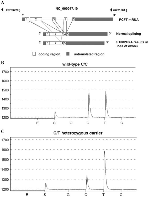
A, The genomic organization and splicing of the PCFT mRNA along with the loss of exon 3 resulting in a truncated PCFT mRNA in patients of Puerto Rican ancestry with HFM. B and C, Representative pyrograms show the wild-type CC sequence and the CT carrier sequence identified from the 1582 newborn samples randomly selected from one year's births (2005) in Puerto Rico. The pyrogram shows a nucleotide sequence around the intron/exon boundary. The y-axis indicates the light emission and the x-axis indicates nucleotide binding. E and S indicate enzyme and substrate, respectively, followed by a negative control base (G), the two peaks of interest (C and T), and a negative control base (C). For the C/T heterozygous mutation, a decrease in the height of the “C” peak and an increase in the height of the “T” peak are compared with the C/C wild-type sequence.