
Fig. 5.
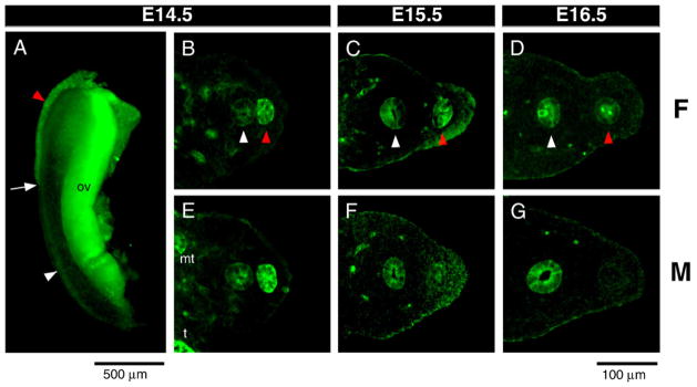
Activated PI3K/AKT pathway is in the Müllerian duct. (A) Phosphorylated-AKT (P-AKT) was detected by whole mount immunofluorescence of E14.5 female urogenital ridge stained with anti-P-AKT antibody. Transverse sections of urogenital ridges at the level of the center of gonads (B–G) stained with anti-P-AKT antibody. E14.5 female (B) and male (E) show that expression in the Müllerian duct is higher than in the Wolffian duct. As the male Müllerian duct undergoes regression (F, G), the intensity of P-AKT progressively decreases to become undetectable. Autofluorescence of blood vessels was observed in the mesenchymal area in B–G. White arrow, tip of the Müllerian duct; red arrowhead, Müllerian duct; white arrowhead, Wolffian duct; t, testis; mt, mesonephric tubules; ov, ovary. Scale bar: 500 μm (A), 100 μm (B–G).