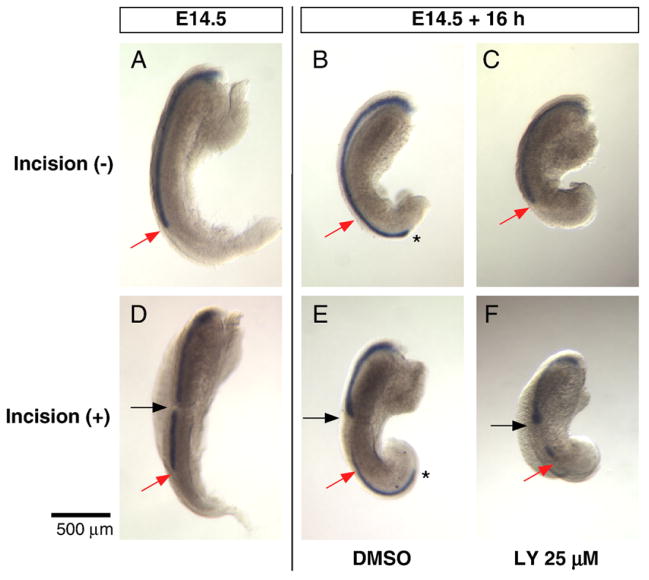
Fig. 7.
PI3K inhibition blocks elongation of the Müllerian duct but not migration of the MDE cells within the incised duct. The Müllerian duct was visualized by whole mount in situ hybridization for Wnt7a (A–F). E14.5 female urogenital ridge before (A) and after 16 h of incubation with DMSO (B) or 25 μM LY294002 (C). The tip of the DMSO treated Müllerian duct reached the posterior end of the urogenital bulge (asterisk in B), but elongation was inhibited as the tip remained at the initial position (red arrow) in the ridge treated with LY294002 (C). E14.5 female Müllerian duct was divided (black arrow in D), and then incubated for 16 h with DMSO (E) or 25 μM LY294002 (F). The tip of the Müllerian duct reached the posterior end (asterisk in E) of the DMSO treated and incised explant; however, elongation was inhibited by the PI3K inhibitor and the tip remained at the initial position (red arrow in F). Despite failure of elongation, the MDE cells have migrated posteriorly (F) and accumulated at the incision site (black arrow in F) and at the initial site of the tip (red arrow in F), creating a Wnt7a blank region. Black asterisks mark the elongating tip of the Müllerian duct. Scale bar: 500 μm.

