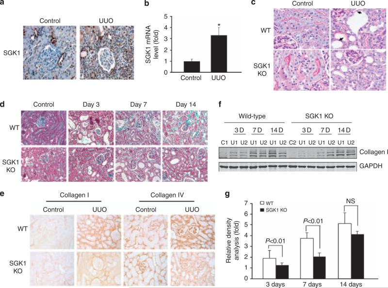
Figure 1. Unilateral ureteral obstruction (UUO)-induced kidney fibrosis was suppressed in serum- and glucocorticoid-regulated kinase 1 (SGK1) knockout (KO) mice.
(a) SGK1 expression in vivo was increased at 3 days after UUO. (b) Real time reverse transcriptase-PCR analysis of UUO-induced SGK1 expression. (c) Kidney sections from wild-type (WT) and SGK1 KO mice with or without UUO for 7 days were stained by periodic acid-Schiff. The arrows point to damaged tubular cells and basement membrane thickening. (d) Sections from control and obstructed kidneys from WT and SGK1 KO mice were stained with Masson's-modified trichrome. The blue color shows extracellular collagen deposition. (e) SGK1 KO inhibited UUO-induced collagen accumulation that was detected by immunostaining with anti-collagen I or anti-collagen IV antibodies and processed with 3,3′-diaminobenzidine (DAB) (brown color). (f) SGK1 KO suppressed UUO-induced collagen I expression. Cell lysates were prepared from control and UUO-treated kidney cortex in WT and SGK1 KO mice after UUO at day 0, 3, 7, and 14. Collagen I expression level was detected by western blot. GAPDH was used as internal loading control. (g) Density analysis of (e). Data represent three repeated experiments. GAPDH, glyceraldehyde-3-phosphate dehydrogenase.

