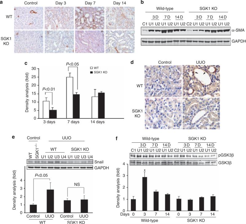
Figure 2. Unilateral ureteral obstruction (UUO)-induced epithelial–mesenchymal transition (EMT) was suppressed by serum- and glucocorticoid-regulated kinase 1 (SGK1) knockout (KO) in vivo.
(a, b) UUO-induced α-smooth muscle actin (SMA) expression was attenuated by SGK1 KO. UUO surgery was carried out in wild-type (WT) and SGK1 KO mice; α-SMA was determined by immunohistochemistry (a) and western blot analysis (b). GAPDH was used as loading control. (c) Density analysis of panel (b). (d) SGK1 KO attenuates UUO-induced Snail expression. Kidney sections were prepared from control and UUO-treated (3 days) kidneys from WT and SGK1 KO mice. Immunohistochemistry was carried out with anti-Snail antibody; the positive cells are shown in brown color. The arrows point to the Snail-positive cells in dilated lumen in kidney tubule. (e, f) Western blot analysis of Snail expression (e) and glycogen synthase kinase-3β (GSK-3β) phosphorylation in UUO-treated WT and SGK1 KO mice. The bar graph shows the density analysis of the western blots. NS, non-significant. *P<0.05 compared with control.

