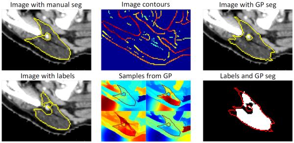
Fig. 2.

Gaussian process segmentation of parotid gland. The initial label from the atlas-based segmentation only partially agrees with the manual segmentation. We extract contours from the image and use them in the kernel function k that allows us to sample label maps , supported by the image. Conditioning these on the atlas labels results in an improved segmentation.
