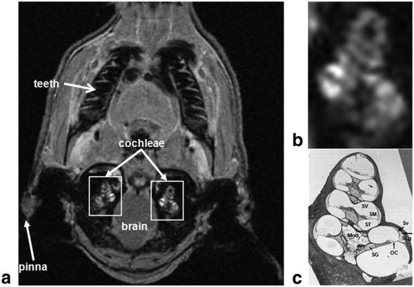
Figure 1.
Location of the cochlea in the guinea-pig skull. a: In vivo proton density MRI (156 μm × 234 μm × 750 μm) obtained 75 min after GBCA injection using a slab-selective 3D Gradient Recalled echo sequence (acquisition matrix=512 × 256 × 16 and FOV=80 mm × 60 mm × 12 mm, TR=20 ms, TE=4.5 ms, FA=10°, 4 signal averages, total acquisition=time 5 min 28 s). b,c: Magnified right white rectangle in a shows cochlear structures (b) identified in a mid-modiolar cochlear histological section (c). c: This panel demonstrates the three fluid-filled cochlear compartments: scala tympani (ST), scala media (SM), and scala vestibuli (SV); the auditory sensory organ, the organ of Corti (OC), modiolus (Mod), spiral ganglion (SG), stria vascularis (Sv), and spiral ligament (SLg). The scala media is filled with endolymph, whereas scala tympani and vestibuli are filled with perilymph. Lateral wall tissues of the cochlea (Sv and SLg) are highly vascularized and characterized by the existence of the blood labyrinthine barrier which allows selective transport of ions, metabolites and nutrients to cochlear tissues.

