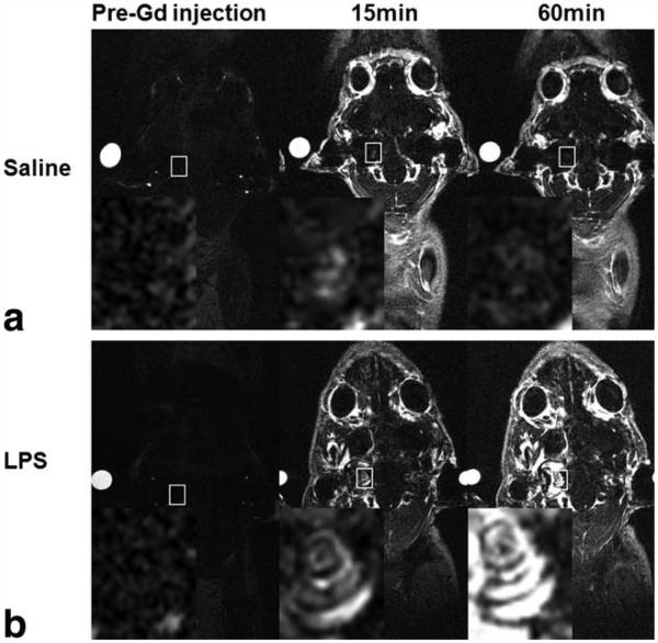
Figure 2.
In vivo Dynamic Contrast Enhanced (DCE)-MRI (156 μm × 195 μm × 750 μm) in coronal orientation acquired after the intravenous injection of GBCA at 1.5 mmol/kg. a: T1-weighted coronal images of cochlear tissues in a control guinea-pig 4 days after saline treatment. b: T1-weighted coronal MR images of cochlear tissues in a LPS-treated guinea-pig 4 days after LPS treatment. Time points indicate the interval after GBCA administration. Reference tube was filled with 5 mM GBCA solution. Inset (magnified white rectangle) shows the marked increase in T1-w signal intensity in the cochleae of LPS-treated animals.

