
Figure 5.
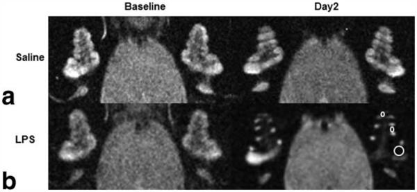
High resolution T2-weighted MR images (117 μm × 117 μm × 700 μm) of the cochleae in control and LPS-treated guinea-pigs 24 h before (baseline) and after (day2) ultrasmall superparamagnetic iron oxide particles (USPIOs) injection. a,b: A control guinea-pig shows a similar signal intensity (a), whereas a LPS-treated guinea-pig shows reduced signal intensity in cochlear perilymphatic compartments in all turns of the cochlea (b, ROIs shown in the left cochlea), indicating the presence of USPIOs 48 h after LPS treatment.