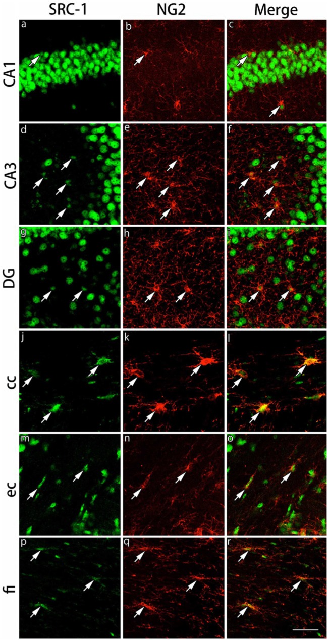
Figure 12.
Double immunohistochemical labeling of SRC-1 and NG2 in the gray and white matter regions. Brain slices were double immunofluorescently stained for steroid receptor co-activator-1 (SRC-1; green) and neuron glial antigen 2 (NG2; red). The staining in hippocampal CA1 (a-c), hippocampal CA3 (d-f) and the dentate gyrus (DG) (g-i) in the gray matter region, and the corpus callosum (cc) (j-l), external capsule (ec) (m-o) and the fimbria of the hippocampus (fi) (p-r) in the white matter region are shown. The cells indicated by arrows show co-localization of SRC-1 and NG2. Expression of SRC-1 in NG2-immunoreactive cells in the white matter regions is stronger than that in the gray matter regions. Scale bar: 50 µm.

