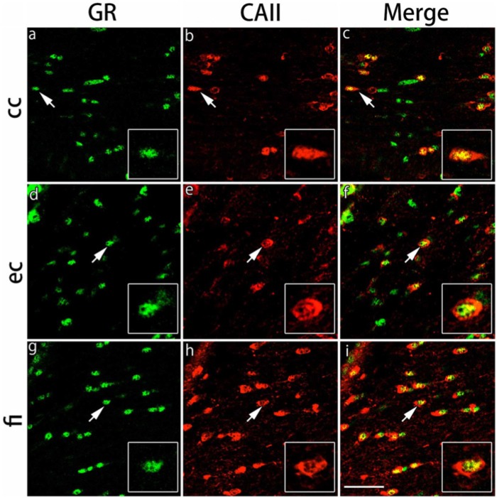
Figure 3.
Double immunohistochemical labeling of GR and CAII in the white matter regions. Brain slices were double immunofluorescently stained for glucocorticoid receptor (GR; green) and carbonic anhydrase II (CAII; red). The staining in the corpus callosum (cc) (a-c), external capsule (ec) (d-f) and fimbria of the hippocampus (fi) (g-i) are shown. The cells indicated by arrows show co-localization of CAII and GR (inset: magnified view). Scale bar: 50 µm.

