Figure 1.
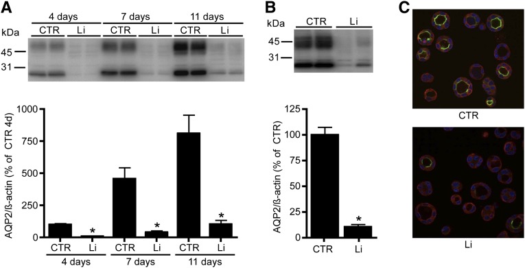
Lithium-induced AQP2 downregulation in two mpkCCD cell models. (A) mpkCCD cells are cultured on transwell filters to study the long-term effect of lithium (Li) treatment. After growth to confluence for 96 hours, cells are treated at the basolateral side with 1 mM lithium chloride and at the apical side with 10 mM lithium chloride. After 4, 7, and 11 days of lithium exposure, cells are collected, lysed, and immunoblotted for AQP2 (upper). Quantification is depicted in the lower panel (n=4 for each condition and time point). (B and C) mpkCCD cells are cultured in matrigel and treated with (Li) or without (CTR) 10 mM lithium chloride. After 3 days, cells are lysed and immunoblotted for AQP2 and signals are quantified, corrected for β-actin (n=4 for each condition). (C) Immunocytochemistry of 3D-grown mpkCCD cells. AQP2 expression is visualized in green, whereas α-tubulin and nuclear 4′,6-diamidino-2-phenylindole staining are depicted in red and blue, respectively. *P<0.05, significant difference from control (CTR).

