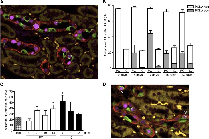
Figure 7.
Lithium causes a G2 cell cycle arrest of principal cells in vivo. Kidneys of mice treated or not with lithium chloride as described in the legend for Figure 6 are isolated, fixed, paraffin embedded, sectioned, and subjected to immunohistochemistry. Simultaneous staining is done for AQP4 (principal cell, red), H+-ATPase (intercalated cell, green), PCNA (nucleus, purple), and pHistone-H3 (G2 cell cycle phase, green). (A) Representative immunohistochemical staining of ISOM area of a mouse treated for 7 days with lithium. (B) Principal-intercalated cell composition (as a percentage of the total number of cells) of the collecting duct of the ISOM of mice treated for 0–13 days (indicated) with lithium and the percentage of PCNA-positive cells in principal and intercalated cells. For each time point, approximately 500 collecting duct cells per mouse for a total of three mice are counted. (C) Percentage of positive pHistone-H3 cells from the PCNA-positive population at days 4–13. (D) Immunohistochemical staining of a kidney after 10 days of lithium treatment. The arrowhead indicates cells that stain for markers of both intercalated and principal cells. *P<0.05, significant difference from the reference value, obtained from a renal cell population of nontreated mice. CD, collecting duct; IC, intercalated cell; neg, negative; PC, principal cell; pos, positive; ref, reference value.

