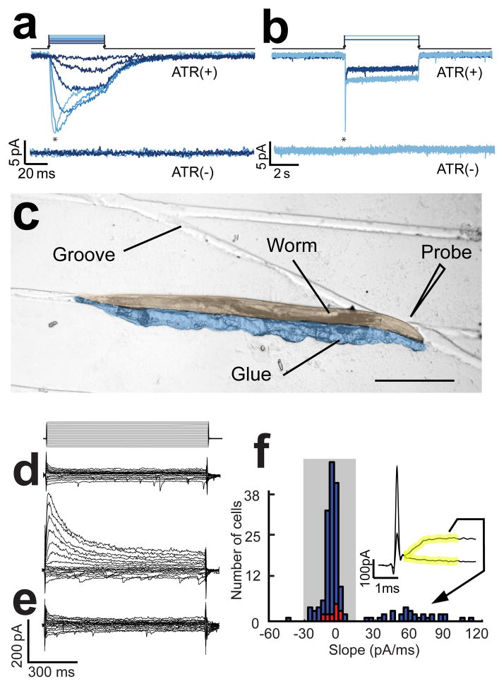
Figure 1.
Photostimulation of ASH neurons and electrophysiological identification of AVA neurons. (a) Membrane current evoked by photostimulation of ASH neurons with 50ms long stimuli; asterisks denote the transient component of the current. The stimulus irradiance varied from 0.07 to 12.5 mW/mm2 in steps of 1.04 log units. (b) membrane current in ASH neurons in response to 5s long pulses of light at 0.75 and 12.5 mW/mm2. Upper and lower current traces are from animals supplied (ATR+) or not supplied (ATR−) with all-trans retinal, respectively. Holding potential was −55 mV. (c) A glued-worm positioned for dissection on a micropatterned agarose substrate. The worm and glue have been pseudocolored to enhance contrast. Scale bar = 200μm. (d) Representatives of the two types of current families obtained when recording from from unidentified command neurons presumed to be either AVA or AVE as identified by cell body position and expression of tdTomato driven by the nmr-1 promoter. Voltage steps ranged from −80 mV to +40 mV in steps of 10 mV from a holding potential of −55 mV as shown above. (e) A current family from an AVA neuron positively identified by combinatorial expression of dsRed and GFP driven by the for glr-1 and rig-3 promoters, respectively. The voltage protocol was the same as in d. (f) Histogram of the slope of the initial current in response the +40mV voltage step in a population of 182 unidentified neurons that were either AVA or AVE (blue bars) and 10 neurons positively identified as AVA by combinatorial expression. Slope was computed over the 2 ms window (yellow) following termination of the capacitative transient ; the inset shows typical traces during this window for the two types of current families shown in d. Shading indicates the subpopulation of neurons identified post hoc as AVA.

