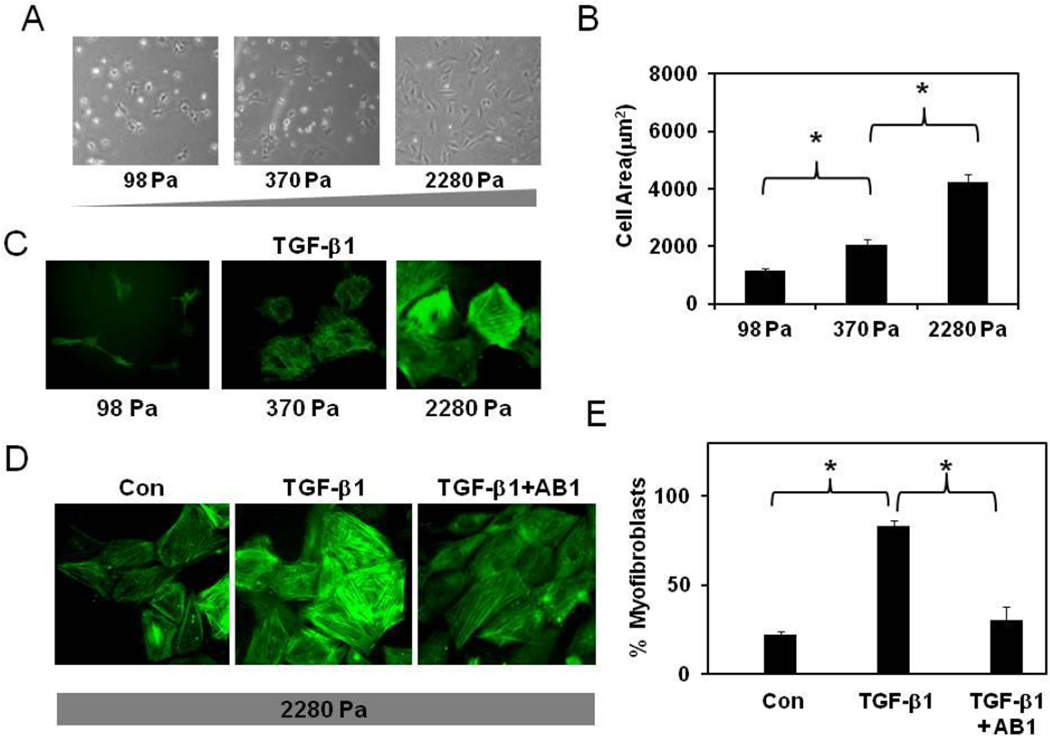
Figure 3. TRPV4 channels mediate TGF-β1-ECM stiffness-induced differentiation of cardiac fibroblasts.
A) Photomicrographs of cardiac fibroblasts cultured on transglutaminase-linked gelatin hydrogels of varying stiffness (98, 370 and 2280 Pa). B) The histogram shows the average projected cell areas of cells (n=200) calculated using Metamorph and Image J software. C) Immunofluorescence images of fibroblast differentiation (the incorporation of α-SMA in to stress fibers (green) on gelatin hydrogels of varying stiffness (98–2280 Pa). Note: CFs were differentiated only on high stiffness gels. D) Immunofluorescence images showing TRPV4-dependent differentiation of CFs on high stiffness gels. CFs were serum starved and stimulated with TGF-β1 for 24 ( C, D ) in the presence or absence of TRPV4 antagonist AB159908 (AB1, D). Cells (n=300) were counted from independent images and the percentage of α-SMA positive cells was presented as % myofibroblasts. The results shown are mean ± SEM from 3 independent experiments (* p < 0.05).

