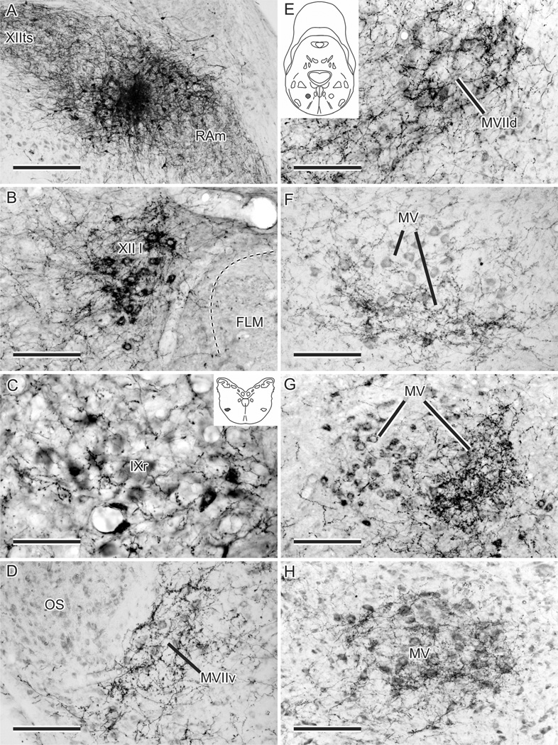
Figure 3.
A–H: Anterograde fiber and terminal labeling in left upper vocal tract motor nuclei resulting from an injection of BDA (A; number 18 in Fig. 2) centered on the right RPcvm, with spread into RAm and intra-axonal transport within RAm and XIIts (see Wild et al., 2009). B,C,G: The motor neurons in XII l, IXr, and MV, respectively, have been retrogradely labeled by CTB injected into relevant muscles (see Materials and Methods). The ventrolateral position of IXr is shaded in the inset in C and similarly for MVIId in the inset in E. Note how the fiber and terminal labeling is concentrated ventrally in MV in F and medially in MV in G but extends throughout much of MV in H. Scale bars = 300 µm in A; 150 µm in B,D–H; 75 µm in C.

