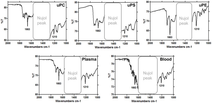
Figure 3. Fourier transformed infrared spectroscopy identifies the crystals produced by different lipids as β-hematin.
The crystals were produced by 100 µM uPC (A), uPS (B), uPE (C) or 10 µg/mL total lipids isolated from PMVM of R. prolixus previously fed with plasma (D) or blood (E). The characteristic iron-carboxylate peaks of β-hematin at 1210 and 1663 cm−1 are shown.

