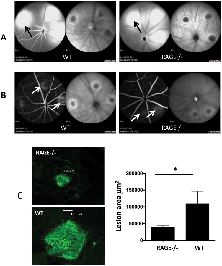
Figure 4. Laser-induced CNV lesions are attenuated in RAGE−/− mice.
Representative images demonstrating laser-burned spots immediately (A) and 7 days after photocoagulation (B) in RAGE−/− mice and WT controls. A) As assessed by cSLO, the retina from WT and RAGE−/− mice shows laser burn sites immediately after photocoagulation. The left image in each pair is fluorescein angiography and the right image is infrared reflectance. There is no obvious difference between the two animal groups with comparable leakage at the lesion (black arrow). B) 7 days after photocoagulation the CNV lesions are apparent in angiograms and infrared reflectance fundus images from WT mice although these are smaller in the retina of RAGE−/− mice (white arrows). C) Comparison of CNV lesion size between the WT and RAGE−/− mice. Retinal flat mounts were evaluated for the presence and size of clearly demarcated isolectin positive CNV lesions one week post-laser injury. RAGE −/− mice exhibited significantly less CNV than age matched controls (n = 12 animals/group, *p<0.05) (Scale bar = 100 µm).

