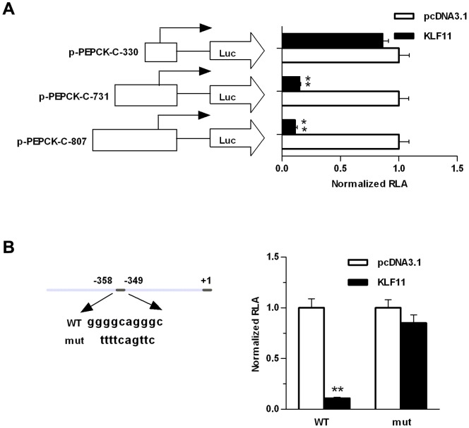
Figure 2. Functional analysis of the promoter region of PEPCK-C in HepG2 cells.
(A) The longer human PEPCK-C promoter construct (pGL3-PEPCK-C-807) and 5′-deleted promoter constructs (pGL3-PEPCK-C-731, and pGL3-PEPCK-C-330) were co-transfected with pcDNA3.1-KLF11 expression plasmid into HepG2 cells, or with pcDNA3.1 (control). After 48 hours, relative luciferase activity (RLA) was measured. (B) pGL3-PEPCK-C-807 promoter construct and mutant construct (pGL3-PEPCK-C-mut) were transfected into HepG2 cells, together with pcDNA3.1-KLF11 expression plasmids or pcDNA3.1 (control). After 48 hours, relative luciferase activity (RLA) was measured. The data shown were the means ± SEM (n = 3). Statistical significance was determined using a two-tailed Student’s t-test (*P<0.05, **P<0.01).

