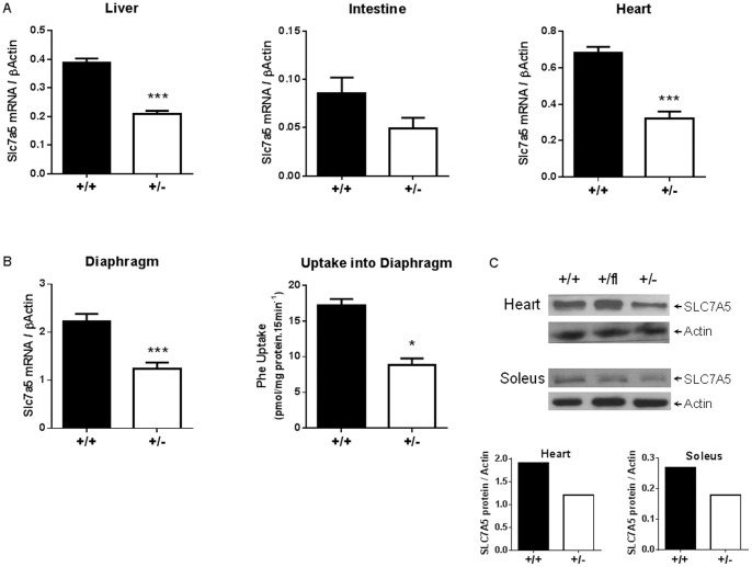
Figure 2. Both Slc7a5 gene expression and SLC7A5 transport activity are reduced in Slc7a5+/− mouse tissues.
(A) Slc7a5 mRNA expression in liver, heart and intestine (as indicated) from Elalpha-Cre Slc7a5+/+ (n = 5–9) and Slc7a5+/− (n = 4–5) mice as determined by qPCR and normalized to β-Actin. Intestine was determined not significant (N.S.) although p<0.1. (B) Slc7a5 mRNA expression in diaphragm from Slc7a5+/+ and Slc7a5+/− mice as determined by qPCR and normalized to β-Actin (n = 6). Uptake of 3H-phenylalanine into diaphragm from Slc7a5+/+ and Slc7a5+/− mice (n = 5). *and ***indicate p<0.05 and p<0.001 respectively by unpaired t-test. (C) Representative Western blot of SLC7A5 protein in heart and soleus muscle lysates from from Slc7a5+/+ and Slc7a5+/− mice. Blot quantitation shown in lower panels.

