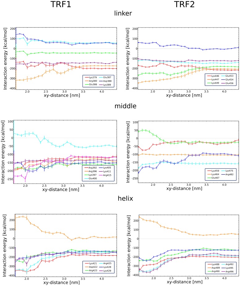
Figure 8. Interaction energy profiles for individual protein residues.
Interaction energy (computed as a sum of electrostatic and van der Waals contributions) between the charged amino acid residues and their surroundings (DNA and solvent combined) as a function of the distance between the protein and the DNA axis. The protein structure is subdivided into three separate regions: the N-terminal linker (top), the middle region (middle) and the C-terminal helix (bottom). For a full set of amino acid interaction energies, see Fig. S7.

