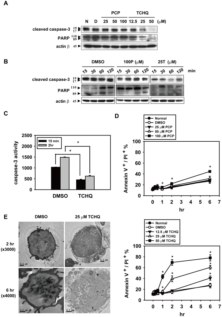
Figure 3. High dose treatment with TCHQ results in inhibition of apoptosis in splenocytes.
(A) Freshly isolated mouse splenocytes were treated with indicated concentrations of PCP or TCHQ for 2 hr. Total protein lysates were analyzed for the expression of cleaved caspase-3 and PARP by western blot. β-actin was served as a loading control. N: normal group; D: DMSO control group. The expression of the target proteins at the indicated time points are shown in (B). Capase-3 activity was detected by a fluorogenic assay, CaspACETM Assay System (Promega, WI, USA), and Caspase-3 released free fluoro-chrome 7-amino-4-methyl coumarin (AMC) was measured at 360 nm excitation/460 nm emission in a Fluoroskan Ascent FL microplate fluorometer (Thermo Scientific, USA). Statistical analysis was performed using a two-tailed Student’s t-test. *, p<0.05. (D) Cell death analysis was performed using Annexin V and PI double-staining. The treated splenocytes were stained for detection of PS with Annexin V-FITC and for DNA content with PI. The necrotic cells were characterized by loss of plasma membrane integrity (annexin V+/PI+). The fluorescence was measured using a FACScan and analyzed by WinMDI software. The statistical analysis was performed using a two-tailed Student’s t-test. *, p<0.05 versus DMSO control group at the time period indicated. (E) Transmission electron microscopy (JEOL JEM-1200EX, Japan) was used to examine cell morphology. Scale bar: 0.5 µm.

