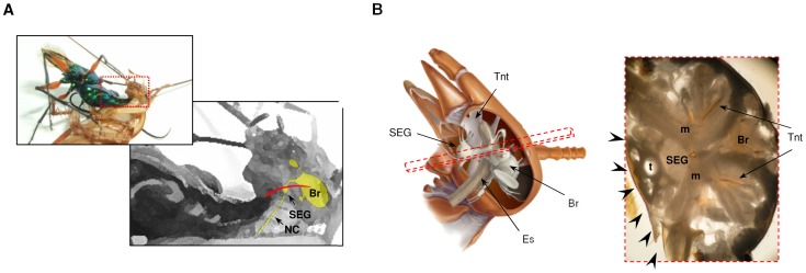
Figure 1. The jewel wasp stings a cockroach into the brain.
(A) A photograph and a diagram showing the presumable trajectory of the wasp’s stinger (red) inside the head of its cockroach host. The wasp holds the cockroach by the pronotum while bending the abdomen towards the cockroach’s head, inserting the stinger through the soft neck cuticle. The central nervous system of the cockroach is depicted in yellow. Br: Brain, SEG: subesophageal ganglion, NC: neck connectives. (B) Left: a lateral view of the cockroach head demonstrating the central nervous system (brain (Br) and SEG), the esophagus (Es) and the internal head skeleton (tentorium; Tnt). Right: light micrograph of a cross section of the head (taken from the plane shown as a dashed rectangle on the left), showing the brain, SEG, internal skeleton, trachea (t) and muscles (m). Different possible points of entry of the stinger through the soft neck cuticle are illustrated by arrowheads.

