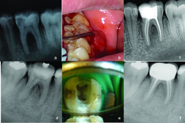
Figure 1.

Case 1- a) Initial radiograph of tooth #36. b) Intraoral film showing increased probing depth values in the furcation area. c) A year recall radiograph showing complete healing. Case 2- d) Initial radiograph showing increased bone lesion in the furcation and distal side of the tooth #46. e) Access opening showing mesio-distal crack line in the pulp floor. f) A year recall radiograph showing healing of the bone lesion.
