Figure 3.

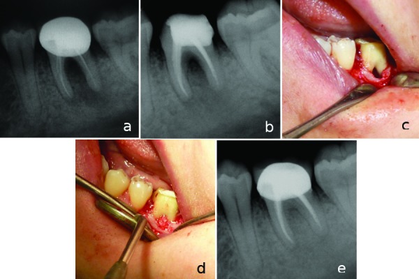
a) Initial radiograph of tooth #36. b) 3 months recall radiograph showing bone lesion in the furcation area. c) Intraoral film showing furcation lesion during periodontal surgery. d) Intraoral film showing bone graft material placement to the furcation lesion. e) Two year recall radiograph showing complete healing of the furcation lesion.
