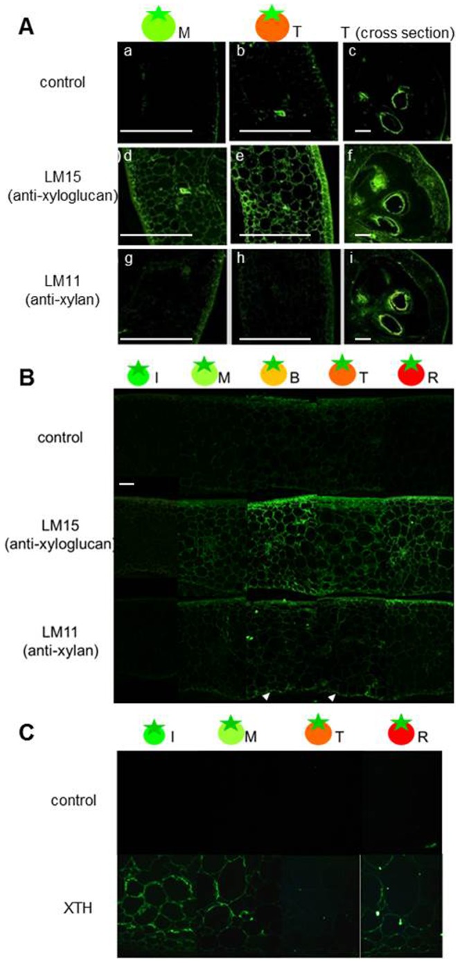
Figure 5. Immunolocalization of XG, GAX, and XTH epitopes in tomato fruit longitudinal and cross sections.

A, Immunolocalization of XG and GAX in fruit sections. Top panels: Negative control (treated with only secondary antibodies). Middle panels: Xyloglucan immunolabeled with LM15 antibody. Bottom panels: Glucuronoarabinoxylan immunolabeled with LM11 antibody. Bars represent 1 mm. B, Immunolocalization of XG and GAX in pericarp. Top panels: Negative control. Middle panels: Immunolabeled with LM15 antibody. Bottom panels: Immunolabeled with LM11 antibody. Bars represent 0.2 mm. C, Immunolocalization of XTH in pericarp. Top panels: Negative control. Bottom panels; immunolabeled with anti-XTH antibody. Bars represent 0.2 mm. Ripening stage: M, mature green; T, turning; R, red ripe.
