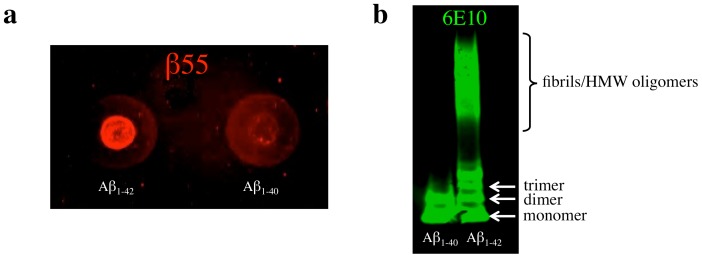
Figure 3. β55 Staining of Dot Blots of Synthetic Aβ Aggregates.
(a) Dot blot of synthetic Aβ1–42 and Aβ1–40 aggregates probed with biotinylated-β55. (b) Western blot of the synthetic Aβ1–42 and Aβ1–40 aggregates probed with 6E10 antibody. The increased staining of Aβ1–42 aggregates in the dot blot relative to Aβ1–40 aggregates is consistent with the greater fibril and high molecular weight oligomer composition of Aβ1–42 aggregates observed in the western blot.

