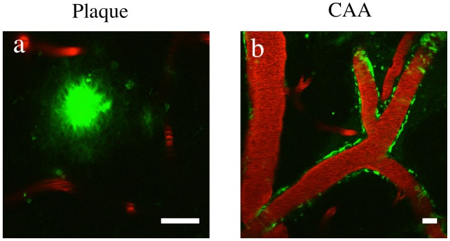
Figure 4. In Vivo Imaging of β55 Positive Amyloid Plaques.
In vivo 2-photon microscopy images from an 18 month old APP/PS1 transgenic mouse obtained 1 hour after topical application of fluorescein-labeled β55 (a,b). Texas Red labeled dextran was intravenously injected for visualization of blood vessels. β55 positive plaques and cerebral amyloid angiopathy are clearly visible in the cortex (a) and vasculature (b), respectively. (Scale bars: 20 µm).

