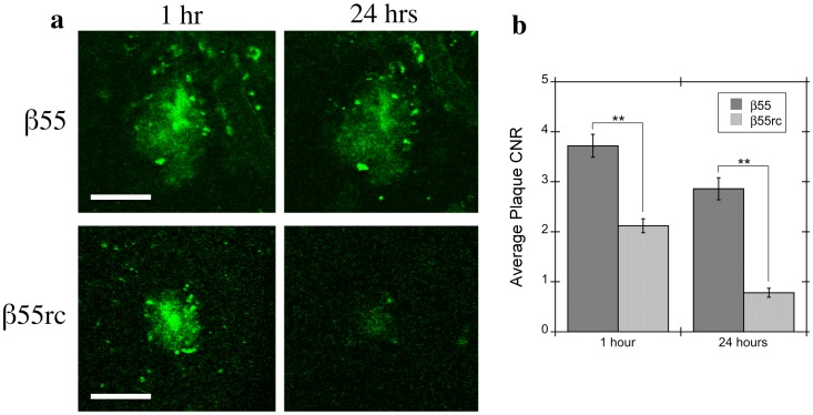
Figure 6. Contrast-to-Noise Ratio for β55 and β55rc Positive Amyloid Plaques.
(a) Representative in vivo 2-photon microscopy images from 7.5 month old APP/PS1 transgenic mice acquired 1 hour (left column) and 24 hours (right column) after topical application of either fluorescein-labeled β55 (top row) or β55rc (bottom row). Most β55 plaques were still visible 24 hours after topical application. In contrast, only a small number of very faint β55rc plaques were still visible after 24 hours. (b) Average plaque contrast-to-noise ratio (CNR) observed 1 hour and 24 hours following topical application of fluorescein-labeled β55 (n = 2) or β55rc (n = 2). β55 positive plaques had a significantly greater CNR than β55rc plaques (p<0.01) at both time points. (Scale bars: 50 µm).

