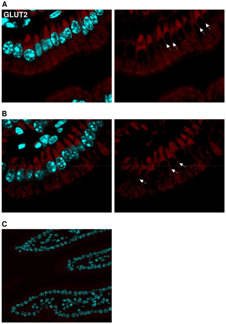
Figure 6. GLUT2 is not located in the apical but basolateral membrane.
Jejunal samples from glut2+/+ animals before and after glucose gavage, respectively, as well as from glut2−/− littermates were stained for GLUT2 (red). Nuclei were stained with DAPI (blue). Basolateral localization (arrows) of GLUT2 in glut2+/+ mice (A) in the basal state and (B) after glucose administration. (C) GLUT2 staining is absent in glut2−/− littermates.

