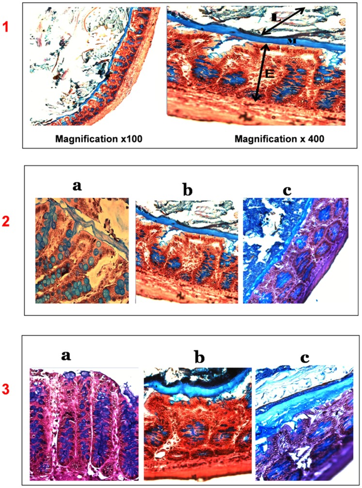
Figure 1. Thickness of the total mucus layer in colonic mucosa in proximal 2) and distal segments 3) of control a), Metronidazole b) and Cotrimoxazole-treated rats c), AB/PAS staining.
1) Magnification x 100 and x 400. The total mucus thickness (M) was measured as a continuous layer between the luminal surface (L) and epithelium (E). Increased thickness of the mucus layer was observed in MTZ and CTX treated rats (b, c). Goblet cells are filled with mucus (blue arrows) either in the control or the MTZ and CTX treated group. Occasionally, a separation could be observed between the mucus layer and the epithelial surface, and may be due to the partial mucus shrinkage during the histological procedure [37].

