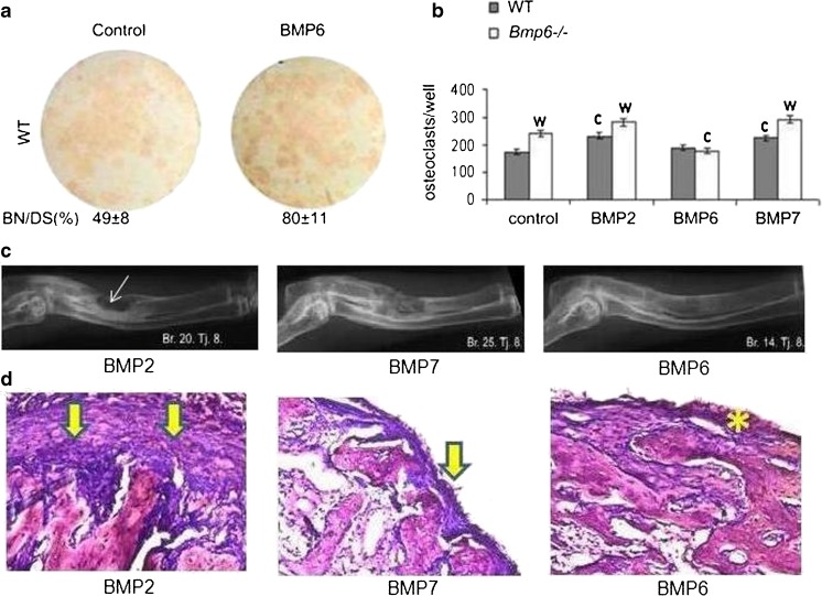
Fig. 5.
Resorption, inflammation/fibrosis and efficacy of BMP2, BMP6 and BMP7. (a) Mesenchymal progenitor cells stained for alkaline phosphatase positive bone nodules which were stimulated by BMP6. Differentiation medium for mesenchymal stem cells added on day 7 contained α-MEM, 10 % FCS, 8 mM β-glycerophosphate, 50 μg/mL ascorbic acid and 10-8 M dexamethasone. The medium was changed every two days until the culture was terminated. BMPs (200 ng/ml) were added to the medium at every feeding. Osteoblasts were identified by alkaline phosphatase stain using a commercially available kit (Sigma Aldrich). (b) Number of osteoclasts derived from HSCs of WT and Bmp6-/- mice. Bone marrow cells were harvested from femurs and tibiae of rats after 14 days of therapy. Differentiation media for osteoclasts contained α-MEM, 10 % FCS, macrophage colony-stimulating factor (M-CSF, 50 ng/mL; Sigma Aldrich), and recombinant human soluble receptor activator of nuclear factor-ĸB ligand (RANKL, 50 ng/mL; Sigma Aldrich). Equal amounts of 200 ng/mL BMP2, BMP6 and BMP7 were added on day 1 and replaced every second day until termination on day 6. The wells were fixed with 4 % paraformaldehyde, and adherent osteoclasts were identified by tartrate-resistant acid phosphatase (TRAP) staining using a commercially available kit (Sigma Aldrich). Significant difference from control (c), and WT (w) (P < 0.05, ANOVA Dunnett test). (c) Efficacy of BMP2, BMP7 and BMP6 in a rabbit critical size defect model. Equal amounts (200 μg) of BMPs were added to 1.5 mL of full rabbit blood, and formulated WBCD was implanted into radius defects of rabbits (n = 4/group. Fourteen weeks following surgery BMP2 implants had a pronounced periosteal rebridgement with endosteal resorption (arrow); BMP7 implants rebridged defects by a cortical union and showed advanced endosteal remodeling; BMP6 treated defects fully healed with advanced bone remodeling and graft incorporation into the radius. (d) Histology of implants (n = 3/group) following subcutaneous axillary implantation of 75 ng of BMP2, BMP7 and BMP6 in WBCD (1.0 mL). BMP2 implant had a thick fibrous capsule on the implant surface (arrows), the BMP7 implant was covered by a thinner fibrous capsule (arrow), while BMP6 in WBCD had no fibrous tissue outside the implant (asterisk)

