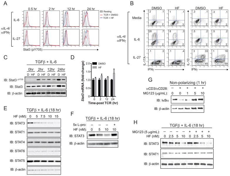
Figure 5. Selective, post-transcriptional regulation of Stat3 expression by the AAR.
(A) Stat3 (Y705) phosphorylation, detected by phospho-specific intracellular staining and FACS analysis, in CD4+CD25− T cells cultured for the indicated times with anti-IL-4 and anti-IFNγ antibodies (αIL-4/αIFNγ) plus either IL-6 or IL-27 alone. Grey shaded peaks – resting T cells (no CD3/CD28 stimulation); red and blue peaks – CD3/CD28-stimulated T cells cultured with vehicle (DMSO) or 10 nM HF, respectively. Data represent 2 independent experiments, each with 2–3 replicates per condition. (B) Intracellular cytokine expression (IL-17A vs. IFNγ – left; IL-10 vs. IFNγ – right) by CD3/CD28-stimulated CD4+CD25− T cells cultured for 4 days with αIL-4/αIFNγ in the absence (Media) or presence of IL-6 or IL-27. Cells were restimulated with PMA and ionomycin in the presence of brefeldin A prior to intracellular staining. Data represent 2 independent experiments, each containing 2–3 replicates per condition. (C) Total Stat3, phospho (Y705)-Stat3, and β-actin protein levels in CD4+CD25− T cells activated for up to 24 hours in Th17-polarizing conditions (TGFβ + IL-6) plus DMSO “(D)” or 10 nM HF, as determined by western blotting. Data represent 3 independent experiments. (D) Mean relative (fold-change) Stat3 mRNA expression ± SD from duplicate samples determined by qPCR in CD4+CD25− T cells cultured for the indicated timepoints (as in (C)). Stat3 expression was normalized to B2M; data represent two experiments. (E) Effects of the indicated doses of HF on Stat protein levels in CD4+CD25− T cells cultured for 18 hours in Th17-polarizing cytokine conditions, as determined by western blotting. Data represent 2 independent experiments. (F) Stat3 and β-actin protein levels, determined by western blotting, in Th17-polarized CD4+CD25− T cells treated with titrating concentrations of HF as indicated for 18 hr. Some cultures were supplemented with 5x (50 mM L-proline (L-Pro)). Data represent 3 independent experiments. (G) IκBα and β-actin protein levels in resting or CD3/CD28-stimulated CD4+CD25− T cells cultured for 2 hours with or without titrating concentrations of MG132. Data represent 2 experiments. (H) Stat3, Stat1, and β-actin protein levels in CD4+CD25− T cells activated in Th17-polarizing conditions for 18 hours in the presence of titrating concentrations of HF +/− 5 μg/mL MG132 as indicated. MG132 was added 12 hours post-activation. Data represent 3 experiments.

