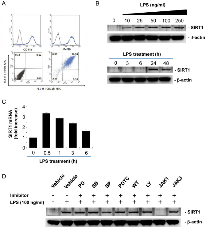
Figure 1. LPS induces SIRT1 expression through JAK-STAT signaling pathway in BMDMs.
(A) Phenotypic characterization of BMDMs by flow cytometry. Mouse BMDMs were analyzed by flow cytometry for CD11b and F4/80 cell surface markers to identify the differentiation of bone marrow cells into macrophages. Over 95% of cells within cultures are double positive. (B) Dose- and time- dependant effect of LPS on SIRT1 protein expression. After BMDMs were incubated with various doses of LPS for 24 h (upper panel) and 100 ng/ml LPS for the indicated times (lower panel), western blot were performed with β-actin as a loading control. (C) Time-dependant effect of LPS on SIRT1 mRNA expression. After BMDMs were incubated with 100 ng/ml LPS for the indicated times, real-time PCR were performed. (D) The effects of signal transduction inhibitors on LPS-induced SIRT1 expression. BMDMs were pretreated with 10 μM PD098059 (PD), 10 μM SB203580 (SB), 10 μM SP600125 (SP), 10 μM PDTC, 1 μM wortmannin (WT), 5 μM LY294002 (LY), 5 μM JAK1 inhibitor (JAK1), and 10 μM JAK3 inhibitor (JAK3), for 1 h and further treated with LPS for 24 h. Full-length blots are presented in Supplementary Figure S1–S2. The Western blot shown is a representative of three independent experiments.

