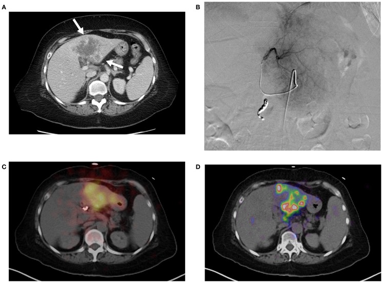
Figure 3.
(A) Pre-treatment contrast enhanced CT imaging of metastatic cholangiocarcinoma in the left hepatic lobe. Areas of increased vascularity (arrow) are seen surrounding areas of necrosis. (B) Left hepatic angiogram during infusion of 90 Y into left lobe. (C) Bremsstrahlung SPECT following radioembolization demonstrates diffuse activity in the region of the left lobe hepatic neoplasm. (D) 90 Y PET/CT demonstrates more detailed information with multifocal areas of maximum activity corresponding to the peripheral viable tumor on pre-treatment imaging.

