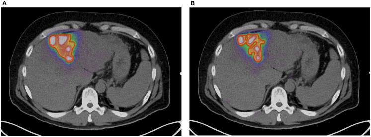
Figure 5.
(A) 90 Y PET of a patient with HCC. Scan performed on Siemens BioGraph mCT Flow with TOF and RR. A continuous bed speed of 0.2 mm/s, 1 iteration, and 21 subsets were used for reconstruction. The contour (red) was performed by an automatic segmentation tool with the lower threshold set to an absorbed dose of 100 Gy. (B) 90 Y PET of a patient with HCC. Scan performed on Siemens BioGraph mCT Flow with TOF, RR, and optimal respiratory gating (HD-Chest). A continuous bed speed of 0.2 mm/s, 1 iteration, and 21 subsets were used for reconstruction. The contour (red) was performed by an automatic segmentation tool with the lower threshold set to an absorbed dose of 100 Gy.

