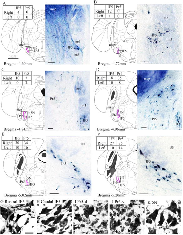
Figure 2.

IF5 labeling after cerebellar tracer injections. A–F: Rostrocaudal series of diagrams and accompanying photographs of labeled cells following a contralateral cerebellar HRP injection (Fig. 1A). The diagrams and coordinates are taken from the atlas of Franklin and Paxinos (2008). The inset tables show the numbers of HRP-labeled cells in the IF5 and Pr5 each side for each section. G–K: Photomicrographs of neurons from each region of interest. 5N, motor trigeminal nucleus; HRP, horseradish peroxidase; IF5, interfascicular trigeminal nucleus; m5, motor root trigeminal nerve; mcp, middle cerebellar peduncle; Pr5, principal sensory trigeminal nucleus; Pr5-d, dorsal part of the principal sensory trigeminal nucleus; Pr5-v, ventral part of the principal sensory trigeminal nucleus. Scale bar = 50 μm in A–F; the scale bar in G is 20 μm, and it applies to G–K.
