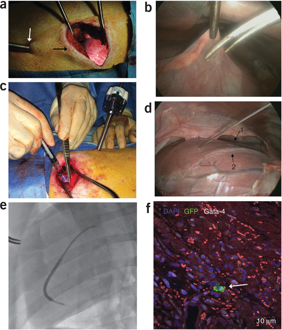
Figure 10.
Surgical- and catheter-based injections of stem cells. Intramyocardial injection model (a) minithoracotomy (black arrow) with thoracoscope (white arrow) shown illuminating the heart. (b) Thoracoscopic view of the pericardiotomy. (c) Intramyocardial injections through the minithoracotomy sight. (d) Thoracoscopic view of the intramyocardial injections. Note the LAD running in the intraventricular groove (arrow 1) and the first diagonal branch of the LAD (arrow 2). (e) BioCardia Morph with deflectable tip directing the Helix biotherapeutic delivery catheter system into the targeted area of the myocardium adjacent to the infarction. (f) GFP+ stem cell identified after intramyocardial injection through postmortem histopathology analysis (white arrow). DAPI, 4’,6-diamidino-2-phenylindole; Gata-4, Transcription factor Gata-4.

