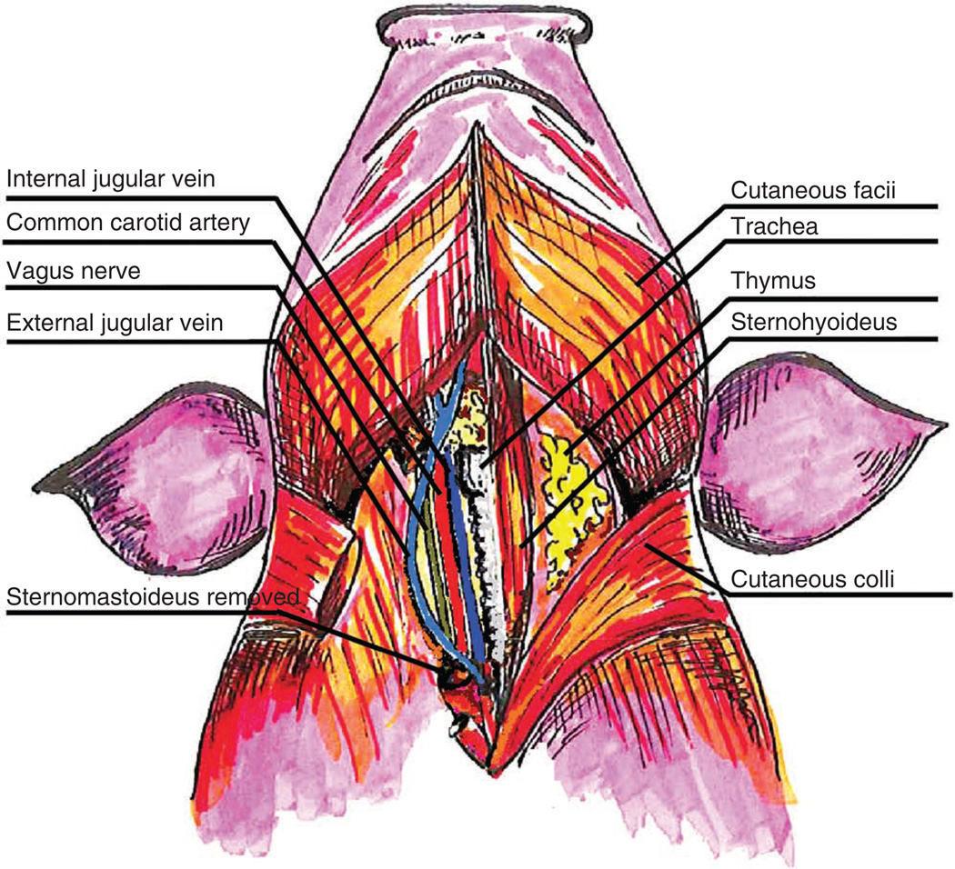
Figure 4.
Neck vasculature and musculature of the swine. Superficial and deep dissection illustrating the landmarks and vessels used for surgical dissection and cannulation of the vasculature in the neck. Note that the sternomastoideus has been removed on the swine’s right to show the underlying anatomical structures in the carotid sheath.

