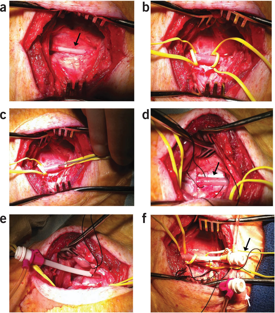
Figure 6.
Vasculature access via a modified Seldinger technique. (a) Right carotid artery exposed and bluntly dissected (arrow). (b) Distal and proximal control of the artery is obtained using vessel loops. Note the dilation of the artery after bathing with 2% lidocaine. (c) 18-gauge, 2 3/4-inch Seldinger needle passing into the arterial lumen to advance an 0.038-inch guidewire before the introduction of a 7F introducer. (d) External jugular vein with proximal and distal 0 silk ties in place for vessel control. (e) 10F introducer in the vein with proximal tie and distal ligation of the vessel. (f) 7F and 10F sheaths in the carotid artery (black arrow) and the external jugular vein (white arrow), respectively, for vascular access.

